As the residency draws to a close, it’s a good time to share images of some of the prototypes and samples that I created alongside undertaking my other research. I always like to document my work and also to be in a position to provide high quality images where necessary, and I can’t do that with the snaps that I take myself. So instead I rely on my wonderful photographer and friend Robyn Manning. Before finishing the residency I took some of the samples and prototypes over to her studio to photograph, and here are the results.
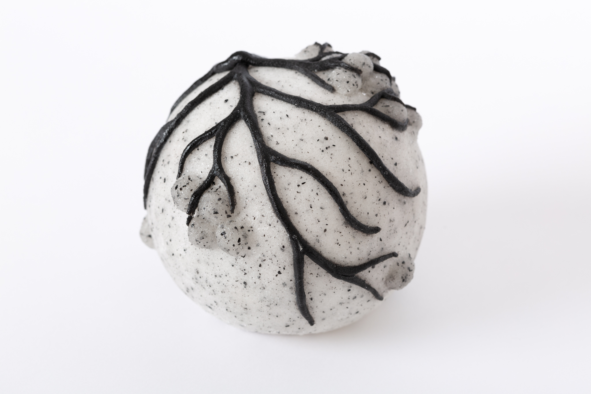
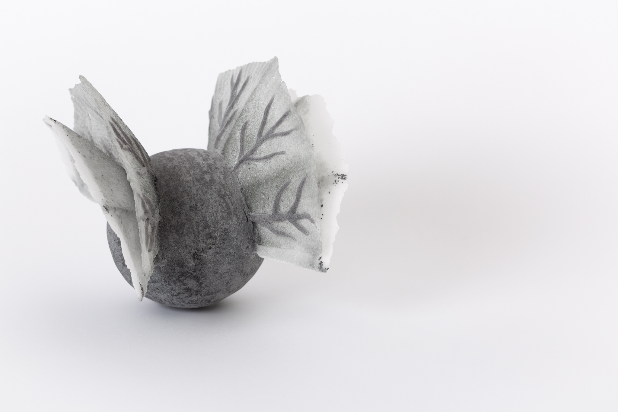
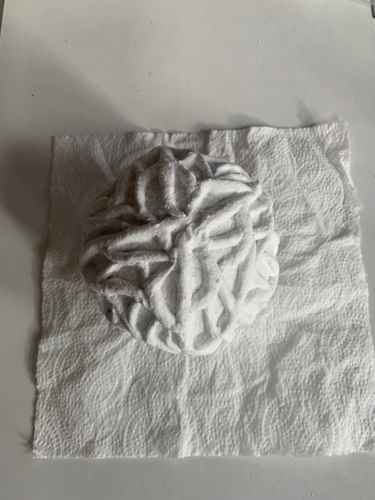
After making the ‘cell’ structured from ‘veins’ I tried a number of different approaches to applying vein-like patterns to the exterior of a cell structure. I wanted to try degrees of visual order or chaos, and to see how these effects looked in different tones – dark veins on pale background, pale veins on pale background, dark on dark etc. I also wanted to assess impact of variations in size. So i made a selection of other ‘dendritic’ cells to compare with the initial cell described in a previous post. Here are some images of the making process for these.
And here are some images of the finished cells
I also made a small sample of a potential flat panel featuring an image of a tangle of blood vessels with gilding added, representing something of a golden thread or pathway through the tangle. I went on to make three larger sample panels which became a series of three with the gilding ‘moving through’ the vein tangle and spreading across the panel.
Panels waiting to be fired for the first time before gilding
Working with different approaches to some of the same theme has felt very productive for me. I have got to assess creatively and technically which types of surface are most successful and most expressive. I also plan to gather some feedback from others as i begin to share the work to get a sense of what the different patterning and finishes convey to those who see them…
I have already written about my interest in angiogenesis as a process that forms part of the cancer ecosystem and that potentially functions as a metaphor in the LCH context (see Concepts and Metaphors (5)). It has been fascinating to look at images of the blood vessels that grow to support tumour growth. and equally interesting to see how clearly related the visual qualities of blood vessels from a tumour are to growth patterns observable around the LCH site, such as amongst the trees and ivy growing near the demokition site and close to the Royal Marsden.
On the left, an image of tumour blood vessels. Centre and right, images of trees and ivy on the LCH site.
These types of images were my starting point for making some glass samples and experimenting with using dendritic growth patterns as both structure for glass cells and for decoration.
And so, angiogenesis has emerged as a focus for my initial creative exploration. I have long been interested in creating vein-like, dendritic structures and decoration, so this is an evolution rather than complete change of direction for my own creative practice.
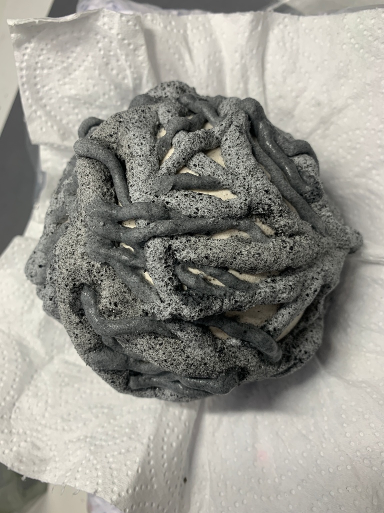
I decided to start with the basic form of the sphere, partly because i enjoy working with that form, and partly because to me the sphere is suggestive of the shape of a cell (regardless of the fact that not all cells are spherical).
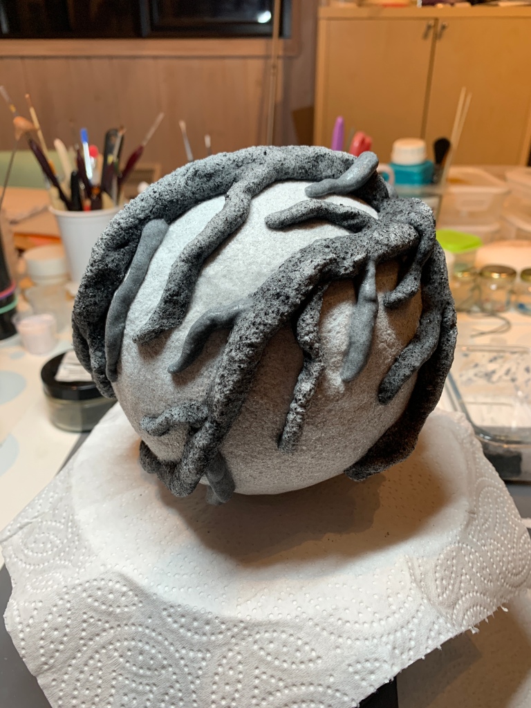
My first idea was to try and construct the form as a whole just from ‘veins’ to see if i could form the cell structure that way. The following images track my progress along that path.
The first line of images above shows the initial ‘veins’ of glass paste that build the structure of the glass over the mould. The second line of images shows a later stage where more veins have been added and interwoven. I was aiming at an outcome that was somewhat evocative of the growth patterns in the images of the tumour blood vessels and also the tree and ivy growth.
In the kiln before firing
This is how the piece looked when it went into the kiln for firing. Glass paste shrinks considerably when it’s fired, so I was expecting the veins to be more slender when they emerged. From the technical perspective I was concerned about whether the structure would be strong enough and stable when it emerged as the shrinkage can also cause the glass to pull away and disconnect or to crack and break. From a creative perspective I wanted to see how the veins looked after firing to see if they (still) evoked the tumour and growth patterns.
Post firing with the mould still insideAfter removing the mould
This is how the finished ‘cell’ looks below with a bit of directional lighting. Luckily the firing went well and the piece emerged intact. As a sample or prototype I am very happy with how it’s turned out. It was very time consuming to make – more so than the samples of dendritic cells that i made subsequently, as the lack of underlying structure for the veins made the whole thing more complex. I also felt that the shrinkage of the glass made the piece slightly less evocative than it was pre-firing, but to some extent this could be changed in subsequent pieces.
One thing I should add at this point is that this – as a sample or prototype – is not intended to be a standalone piece. My intention was that it could be a model for a component of a composition of cells, potentially some in light, some in shadow. I knew also that I wanted to experiment with working in a pale colour, probably white, and making vein structures that were also more ordered, less chatotic. So that’s what I did next.