A few weeks ago I was lucky enough to meet with the PPIE team at the Royal Marsden. Their role is public and patient involvement, and they do an amazing job to engage patients and local communities with the work of the hospital. I had a great conversation with them about their work and also a really constructive conversation about how I might be able to involve some current or former patients in my project.
The immediate suggestions were that I have a page on their platform the Cancer Patients’ Voice and that they approach the patients on their database to see if anyone would be interested in being involved. These are both ways they habitually give patients the opportunity to join research or other activities at the hospital where patients’ input is crucial. And while my project is no way typical of the kind of thing that normally features, I was grateful that they didn’t see that as a problem – in fact they were enthusiastic about having a different sort of activity that patients might be interested to contributing to.
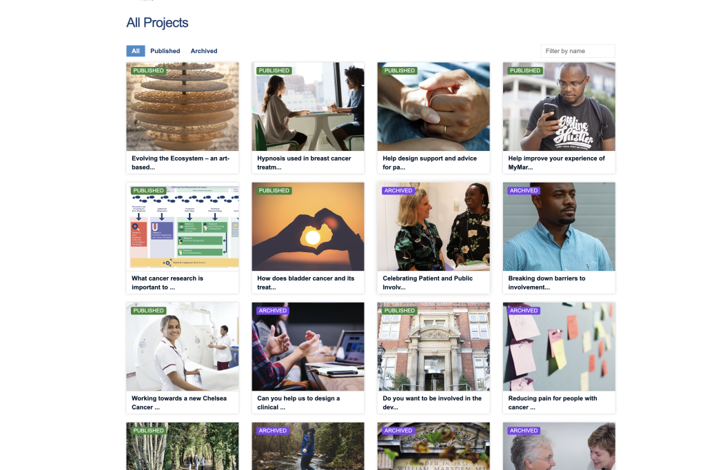
These images above show my page as it was published at the beginning of November. I’m also delighted that I’ve already had contacts from a number of current patients who are interested to find out more about being interviewees for the project. I am delighted as it feels llike the patients’ point of view is an critical part of understanding the ecology of cancer treatment.
Today’s ideas are all about routes, travelling, and journeying. Some seem to be so commonplace that they are not really thought about as metaphors at all. And I’m also beginning to pick out metaphors that are more clearly ‘metaphorical’ and used deliberately because of their non-scientific connotations.
Concepts in a cancer context
Many of my descriptions about how these relate to cancer are drawn from a fascinating conversation with the Biology of Childhood Leukaemia team in a discussion about the role of gene TP53 in regulating cancer and the effects of hypoxia (lack of oxygen) on cancer evolution.
“Pathways”
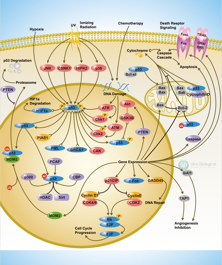
Steps in the process that govern a cellular system. For example, the TP53 pathway is the one that a cell goes through in terms of whether it is ‘allowed’ to replicate, is sent to be fixed or if it is earmarked to die.
An example of how a pathway is illustrated – this one centred on the P53 protein generated by the TP53 gene
“Gateway”
On a pathway there can be a gateway, such as the gateway that cells go through on the TP53 pathway, for example, in order to know if they should go forward to replicate or stop and die. This is a gateway that can become very ineffective in cancer – cancer cells do not die in the same way as normal cells – and may be related to changes or mutations of the TP53 gene.
“Bottleneck”
A point at which many cells fail and a few pass through such as in the toxic environment at the centre of a tumour where the majority of cells die but one or two may replicate with mutations that allow them to survive.
“Signalling”
How cancer cells communicate with surrounding cells. Eg, they can signal to the body to grow more blood vessels to a tumour. Signals can be proximal (ie next door with surface proteins) or longer range chemical (eg with hormones etc).
“Rite of Passage”
This is a more specific metaphor drawn from one of the papers of the Childhood Leukemias Team. This metaphor represents the point of no return in cancer. Specifically, the ‘rite of passage’ in the paper refers to the the point at which the TP53 mutation allows cells to transition through EMT and launch themselves into the rest of the body. THis is the point where cancer metastasises, after which the hopes of ‘cure’ are drastically diminished. A rite of passage indeed.
How all this relates to the London Cancer Hub
While talking to ICR researchers the idea of spatial movement came up frequently, both in terms of their research and about their personal movements around the site and around London. While it seemed to me that in terms of the site, they concentrated on the ICR part of the campus, several mentioned how walking past or through hospital buildings either as part of their journey to work, or to collect samples or meet with clinicians, reminded them of the ultimate purposes of their work. This was especially true when they cam into direct contact with patients, even if this was just seeing or passing them on their own pathways,
Movement around the LCH was not the only example of establishing pathways in a very literal sense. Several researchers also told me about their regularly trodden routes around London as part of their work. For example, one scientist talked about how at one stage in her work she was travelling frequently – occasionally daily – on a circuit between the Sutton ICR, the Chelsea ICR and the Imperial College campus at White City. Interestingly, her work was heavily focused on identifying the spatial arrangements of different cell types in tumours, which are ‘barcoded’ to keep track of their position. We chatted about the possibilities of tracking the movements of researchers in a similar way. I would love to track some staff across the site and see what visual mappings came out of the exercise. I’ll be posting more about the site, its history and pathways through it in a forthcoming post…
And the ‘rite of passage’ for the LCH? I could interpret that in so many ways, so I am going to hold back and see what else emerges,
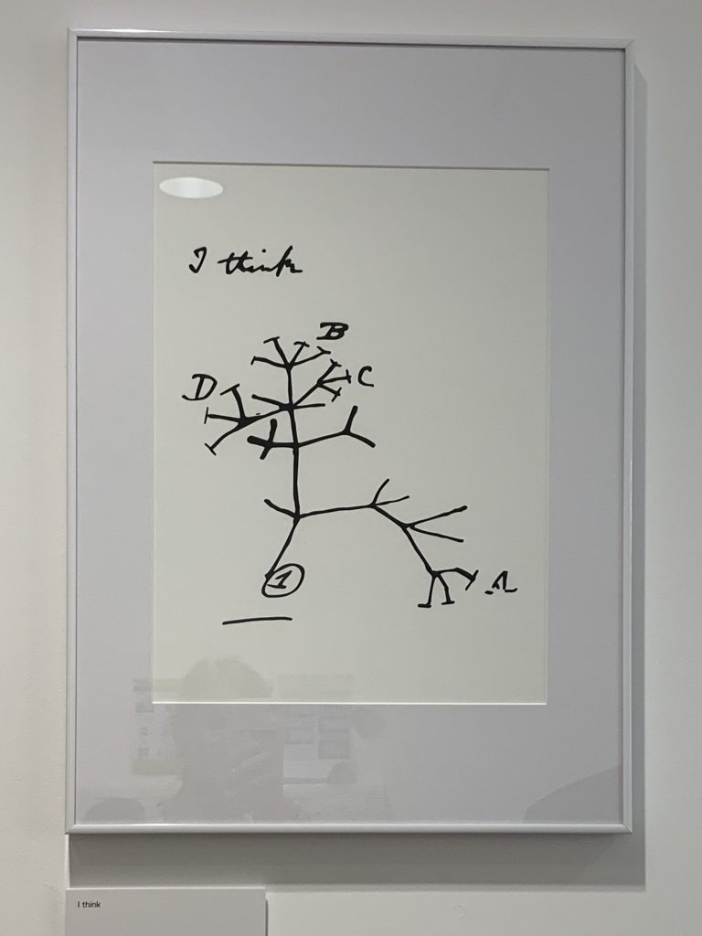
One of the initial priorities for my ‘Evolving the Ecosystem’ project was to learn about some of the key ideas and concepts that underpin the study of cancer as an ecosystem. I also want to get to grips with how they might be applied to the work of the teams at the LCH. So my first series of meetings at the LCH have been primarily focused on researchers at the Institute of Cancer Resarch (ICR) who have generously explained the thinking behind their research, sharing ideas and research papers and painstakingly explaining complex biology to a non biologist. First off, I am very grateful to all who met with me from the Biology of Childhood Leukaemia team and from the Centre for Evolution and Cancer teams and who spent time with me on my visits in late September and early October. I am not planning to record the content of each of the conversations here, just to say that they were incredibly illuminating and introduced me to a wealth of ideas around cancer ecosystems.
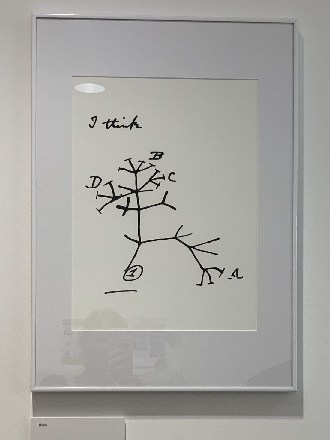
Darwin’s ‘I think’, mounted outside offices at the ICR
From these discussions I have started to draw out some of the concepts that I think may be useful going forward to inform my artwork. Scattered through future posts I will be sharing selections of the concepts and metaphors I am encountering, with my interpretation of their meaning in the context of cancer, in the context of organisation, or better still, both. I would emphasise here, though, that all the interpretations I am sharing are mine. They may be drawn from conversations with researchers and others but if they are full of errors or misunderstandings, those are entirely my own responsibility.
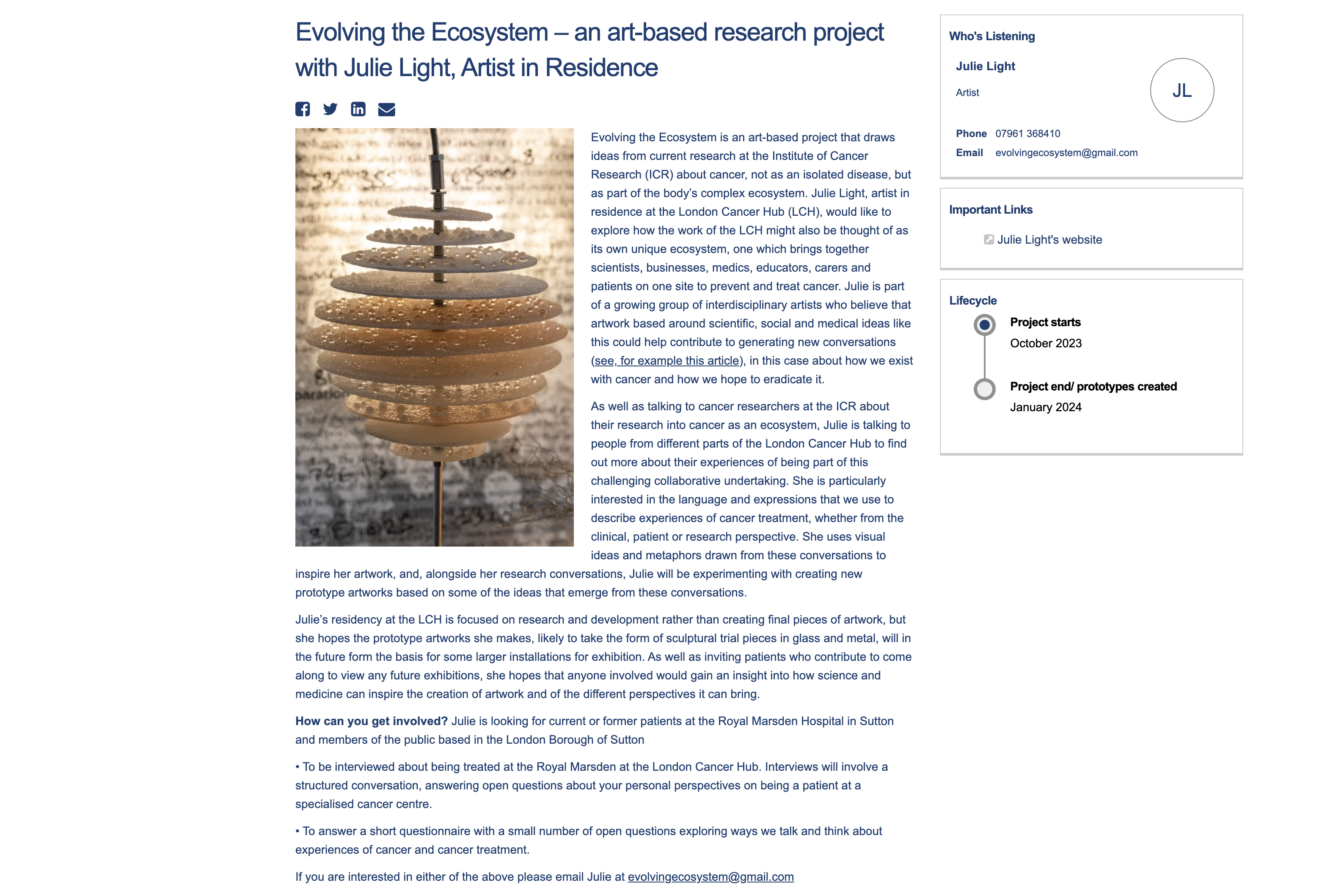
My residency at the London Cancer Hub will be spent working on a project I have called ‘Evolving the Ecosystem’. A central goal of the Institute of Cancer Research (ICR) strategy is to unravel the cancer ecosystem; cancers develop and grow very much as part of the environments of our bodies, and a priority for research at the ICR is to understand the mechanisms by which our individual or common internal bodily ecosystems govern whether cancers thrive or fail.
I was inspired by how the concepts we draw from ecology affect not only how a disease progresses, but potentially also how organisations thrive or fail in response to their environments. Below is an extract of the project proposal I put together to explain the research that I want to do whilst on this residency.
“Cancers have increasingly been seen as part of the complex ecology of the body rather than as diseases where rogue cells or tumours can be understood in isolation. Research scientists and clinicians are now focusing on investigating the characteristics and mechanisms of this ecology to find new ways to control or eliminate cancer. The conceptual framework of the ecology of cancer – and how to unravel it – has proved so useful that it now forms a central plank of the Institute of Cancer Research’s (ICR’s) current strategy.
Organisations too can be understood as ecologies. The London Cancer Hub (LCH) brings together a cross section of organisations working towards a synergistic set of goals, forming its own complex ecosystem, an ecosystem aiming to create an impact which is greater than the sum of its parts. How the constituent organisations – involving research scientists, businesses, clinicians, educators, carers and patients – collaborate across the new Hub to achieve this can similarly be viewed as an endeavour to develop a healthy ecosystem.
For this project I want to investigate these parallel ecologies side by side, drawing on frameworks and metaphors usually confined to the scientific context to explore what we can learn about the development of a culture and environment within which the potential for research, treatment and prevention can thrive.
The artwork emerging from the project would act both as an expression of the learning from the project and as a stimulus for further conversation going forward about how to build our capacity to tackle cancer. In addition, the stories gathered along the way would capture a moment in the history of cancer research and in the development of the LCH”.
I hope this conveys some of my ideas behind the project – which will surely also evolve as the project progresses. And if you have any thoughts about this that you’d like to discuss, do get in touch…..
I am delighted to say that I shall shortly be starting a new project, another collaboration about which I am very excited, so I will be posting again regularly to record my research and learning as I create and develop a new series of artwork. Watch this space to hear more in the very near future!
So, for those of you who have read my post on placing the artwork, you know that there are some major advantages to placing the artwork under glass (even if it is itself made of glass!).
I know that I don’t just want to make an artwork and pop it into a standard cabinet – often glass art does not fare well when placed inside a glass case. So my plan is to create an artwork where the cabinet or vitrine is an integral part of the piece.
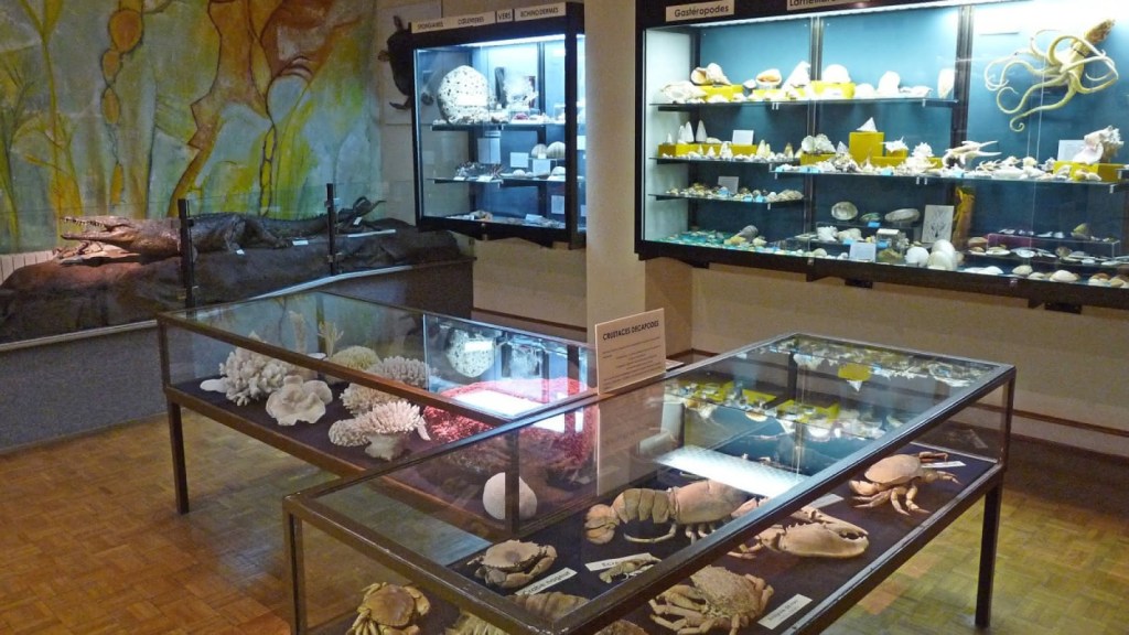
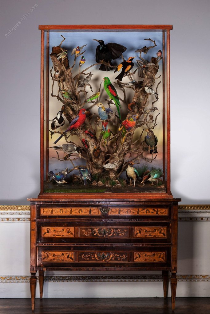
When thinking about how to use cabinets or vitrines in this artwork, I am very much drawn to thinking about natural history exhibits. A lot of natural history museums in particular evolved complex settings for displaying their flora and fauna specimens in their vitrines during the late nineteenth century and enduring well into the twentieth century. This approach also spilled over into more general taxidermy.
Taxidermy display of Australian birds
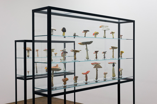
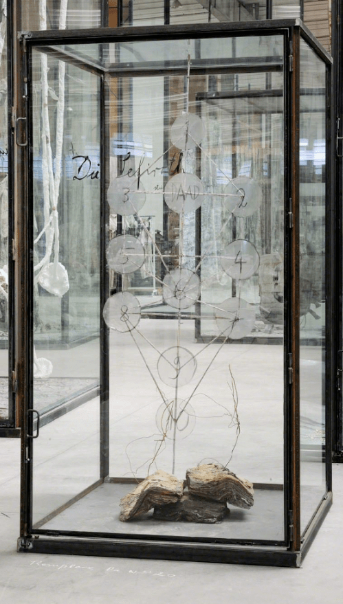
Various artists are also known for using vitrines in their work. Here are examples from Carsten Holler, Anselm Kiefer, and an artist new to me Fiona Hall. They have used vitrines in different ways, creating types of taxonomy, mises en scenes and
Carsten Holler’s meditation on chanterelles and the colour orange.
Anselm Kiefer
Anselm Kiefer
Fiona HallFiona Hall
Reviewing how curators, collectors and artists use of vitrines, some of the key things I know I want to think about going forward include:
Backdrop – coloured and or sandblasted
Drawing or writing on the case
Mise en scene inside the case – including narrative elements
Composition of main elements as specimens (or not)
Integrated lighting
I already have my eye on a specific vitrine / display case so am going to be thinking about those things in light of that….
If you haven’t already read my account of days one and two, you might like to scroll down and start at the beginning of my trip. If you have, or like to start things in the middle, read on!
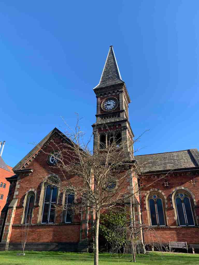
Day three of my visit saw me heading for St James University Hospital in Leeds, rather than the University. After I walked past a very picturesque Victorian chapel and restored Tom met me in the Wellcome Trust Brenner Building. This University building on the hospital campus houses many of the Biology researchers and their labs.
The Victorian Chapel at St James’ University Hospital in Leeds – rather more picturesque than the Wellcome Trust Brenner Building where i spent much of the day.
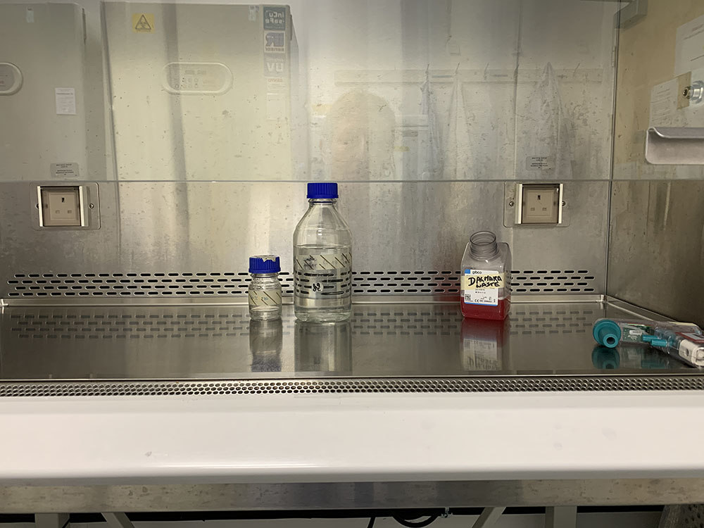
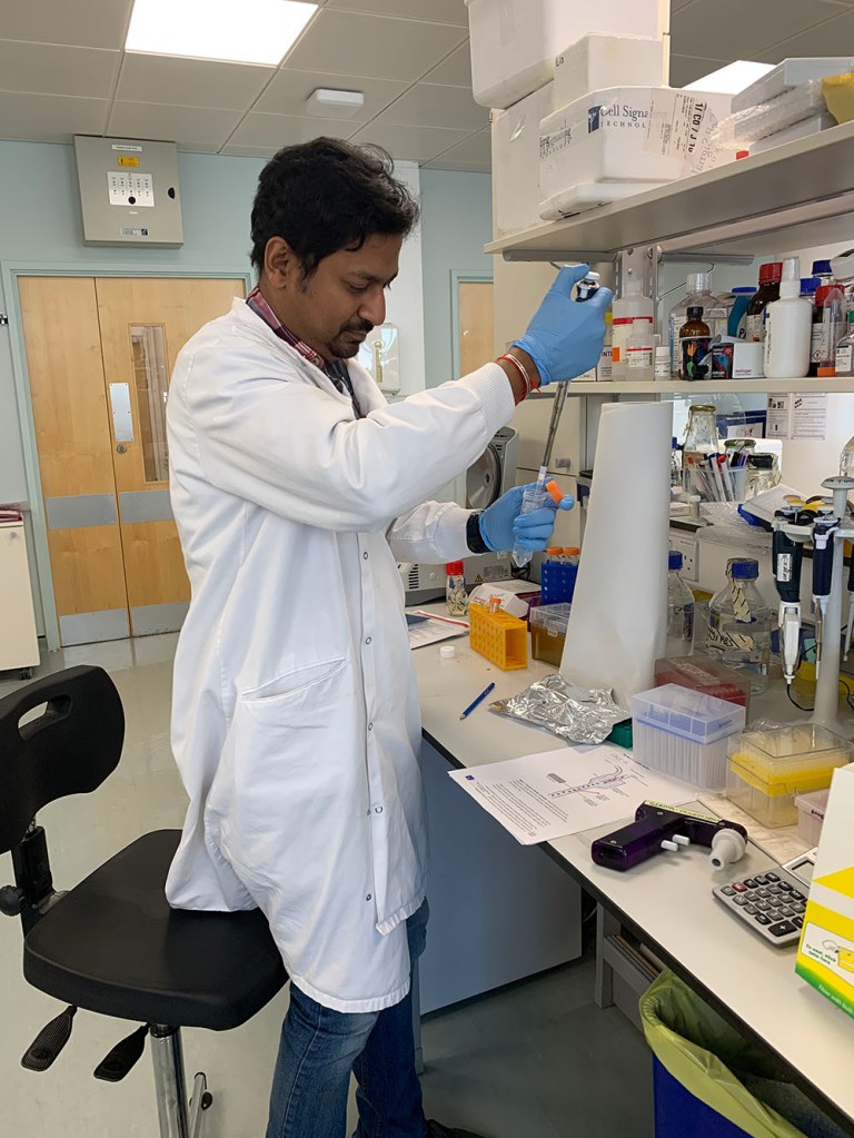
After a lightening tour of the labs and offices, Sarah gave me an induction about the basics of lab safely and etiquette for my role as an observer. I learned where and when to wear a lab coat, what not to touch (basically everything in the lab, including not leaning or propping my notebook on any lab surfaces), when to wear gloves and when to wash hands. With all the current advice, I have already got a lot more skilled at handwashing, so that stood me in good stead.
Arindam prepping samples
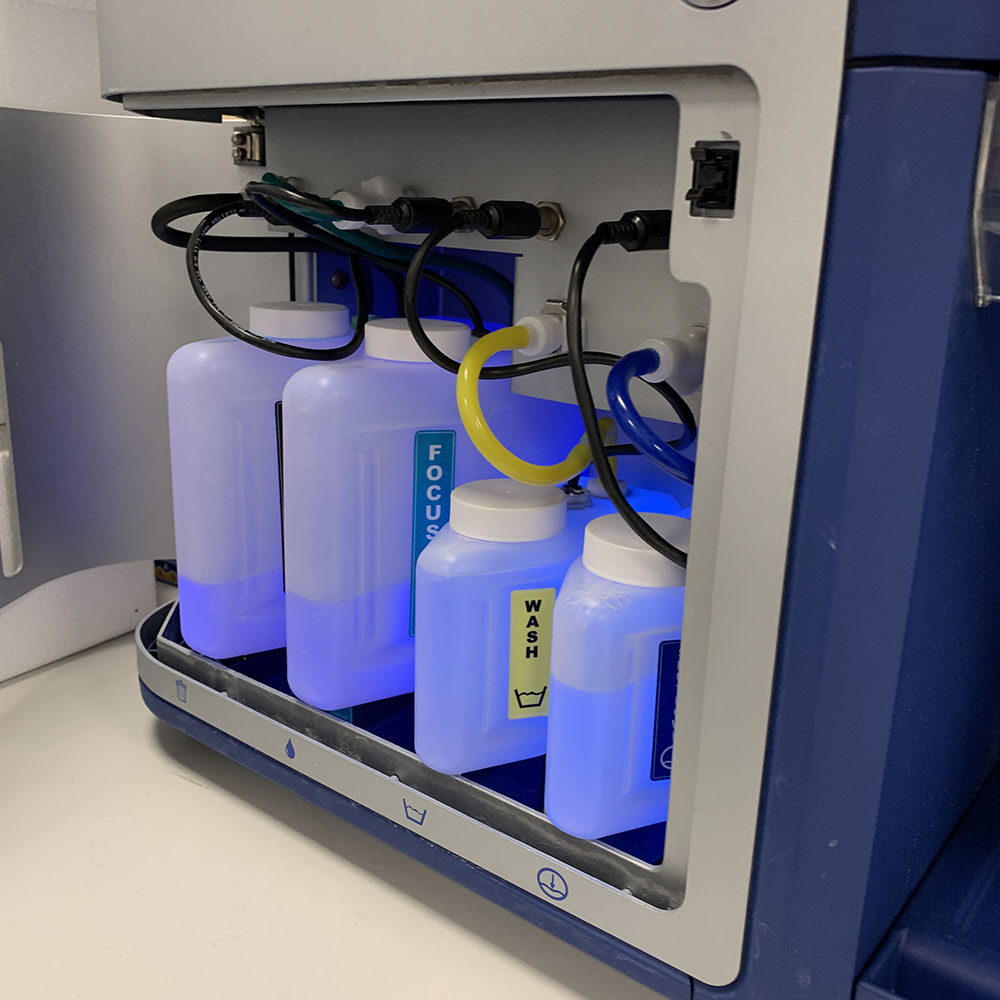
After that, I was handed into Arindam’s care. He had been prepping some experiments that we could run through the flow cytometer, part of the three day protocol that he had started before I even arrived in Leeds. The flow cytometer is an instrument which can differentiate very quickly between different cell states – which are tagged with different fluorescent labels – by passing them one by one through a narrow channel, shining a laser at them and assessing their luminosity, before chucking them back into the main sample. Amazing. Oddly, it made me think of counting sheep by making them jump over a gate.
Here are the innards of the flow cytometer – a symphony in blue.
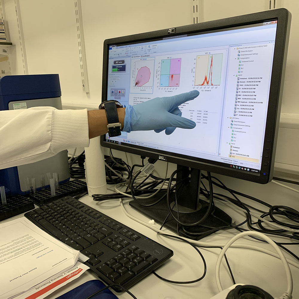
The read out from the cytometer is shown in a graphic display on the cytometer’s monitor. Here we are looking at the percentage of cells that have died as a result of being treated with the MP1 peptide. The four box model on the screen shows living cells, early apoptosis, late apoptosis and necrosis after treatment with a particular peptide concentration.
The team are regularly working with a number of different cells line. There are four in particular that the team are using as their ‘core’ cell lines. Three of these are different breast cancer cell lines, and one is a ‘normal’ cell line (although modified so that it will continue to grow in a lab). I am really interested in how the cell lines were chosen and some of the complexities and ethics of working with cell lines, so expect to hear more about this in a separate post. My initial reaction, though, to the normal-but-immortal cell line is this: if what distinguishes a normal cell from a cancer cell is that is a cancer cell will grow and reproduce without being properly programmed to die, then the existence of a normal-but-immortal cell line is something of a conundrum – not a total contradiction but not straightforward either.
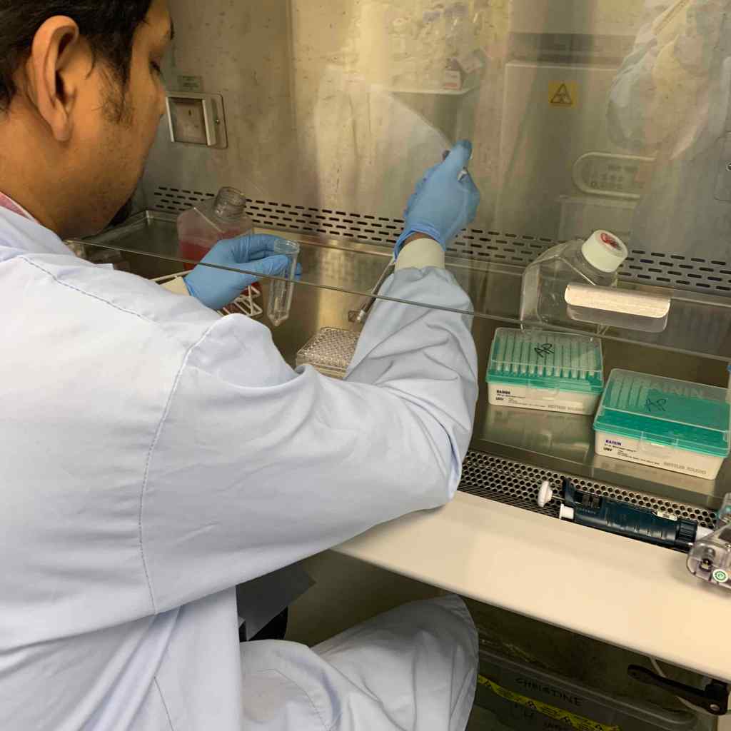
Dosing cells with MP1
Later in the day Arindam showed me some spheroids to be treated with the peptide which we could then view through the confocal microscope to see how the peptide affected cells in a 3D configuration. In the previous experiment, the cells had been standalone, but if you are to treat cancer effectively, you are much more likely to have to treat clusters of cells. Using the normal-but-immortal cell line, Arindam had prepared the spheroids of clusters of cells – apparently the cancer cell lines don’t make good spheroids, just random bunches of cells that aren’t useful for testing.
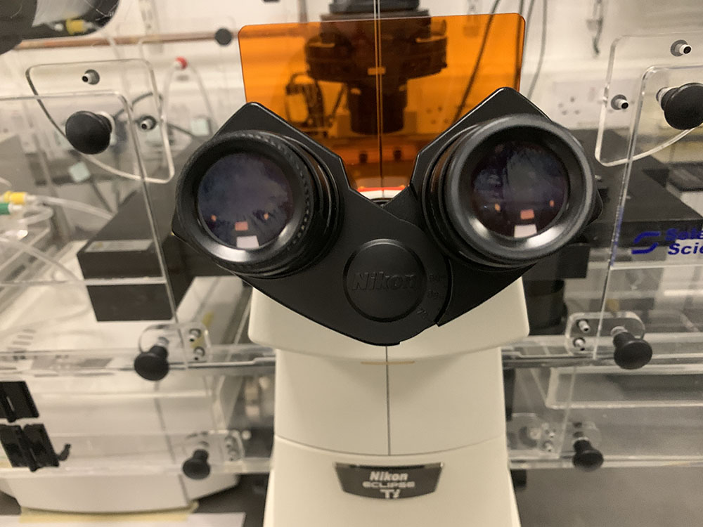
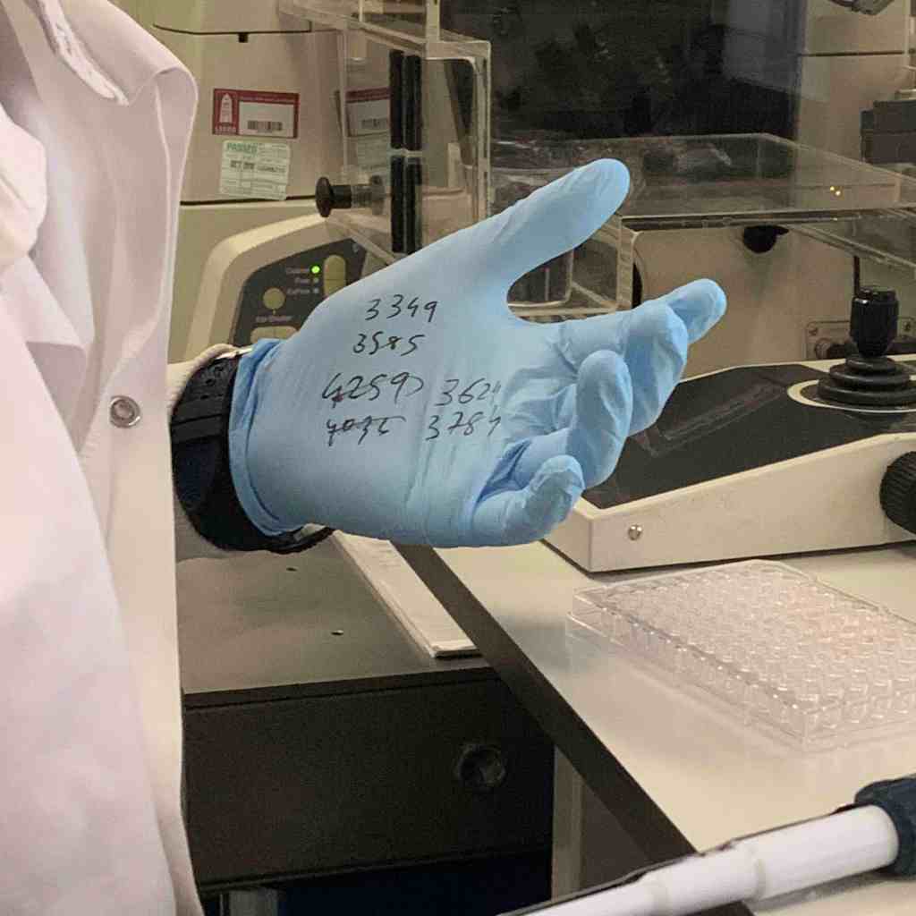
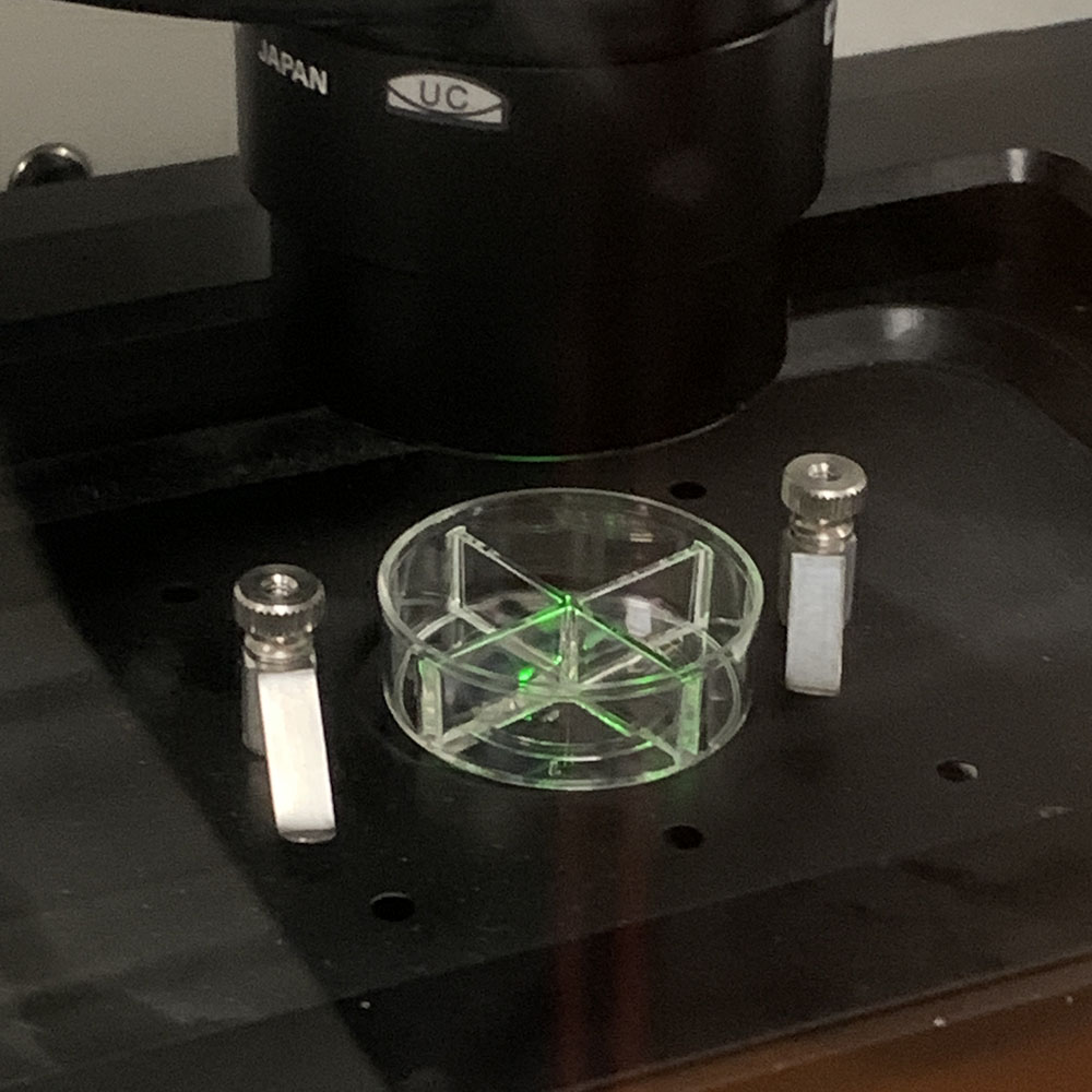
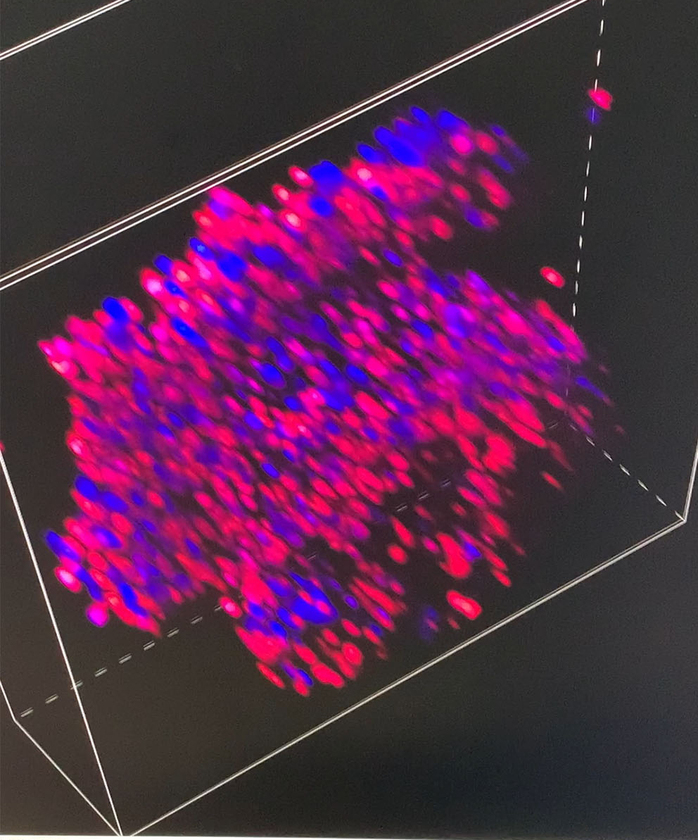
The confocal microscopeWhen the easiest place to record your calculations for the ‘z stack’ is your gloveThe illuminated spheroid sample sitting in place under the microscope lensAn image captured from the animated z stack sequence – very low res as we didn’t have time to allow it to process a higher resolution version
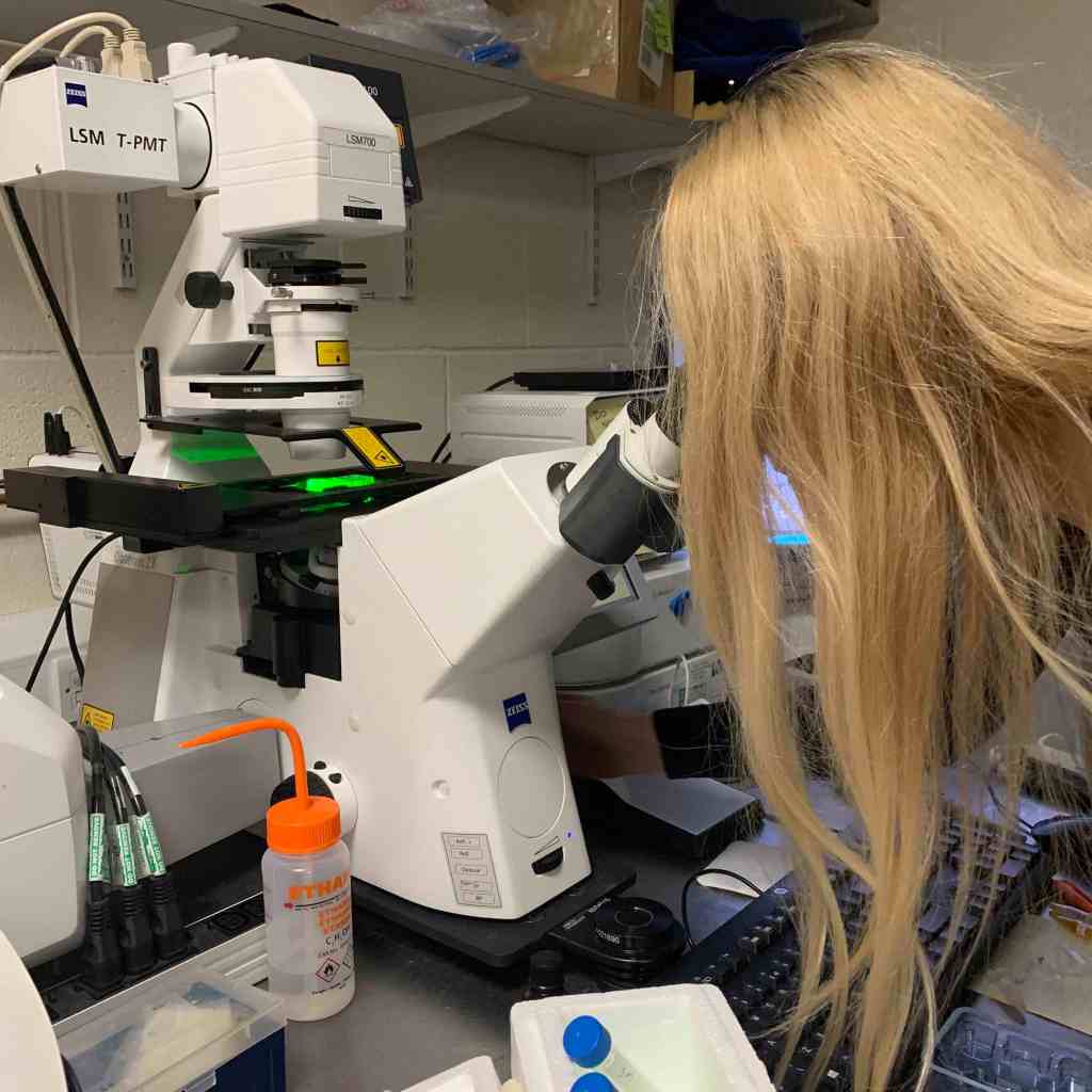
After Arindam and I had looked at various spheroids treated with different amounts of peptide, I went to spend some time with Dagmara. Using a simple optical microscope, we looked at samples of GPMVs that she had been creating from all of the different cell lines.
Dagmara and the microscope.
Dagmara is studying for her PhD as part of the project. One of her priorities so far has been to develop ways to create GPMVs that can be used as part of the peptide testing process. GPMVs are, to me, like a halfway house between testing on GUVs (or LUVs) and testing with actual cell lines. A GPMV is a vesicle created from an actual cell, so the membrane has the same (or similar) composition in terms of naturally occurring lipids etc that the cell has. It is therefore a much more complex membrane mixture than a GUV. But it doesn’t contain all the active gubbins of a real live cell, so there are fewer variables with testing GPMVs than there are with real cells.
However, it turns out that creating GPMVs – which are Giant Plasma Membrane Vesicles – are not easy things to create. Dagmara has been using two approaches – oscillation and chemical. Both techniques are tricky and oscillation only seems to work with one cell line. And even where she has been making GPMVs successfully, there are problems with them being ‘leaky’. This is a proper problem as the action of the peptide – as i understand it – is to create pores in the membrane that creates leakiness and causes the cell to expire. If you start with a leaky vesicle, that’s hard to test for.
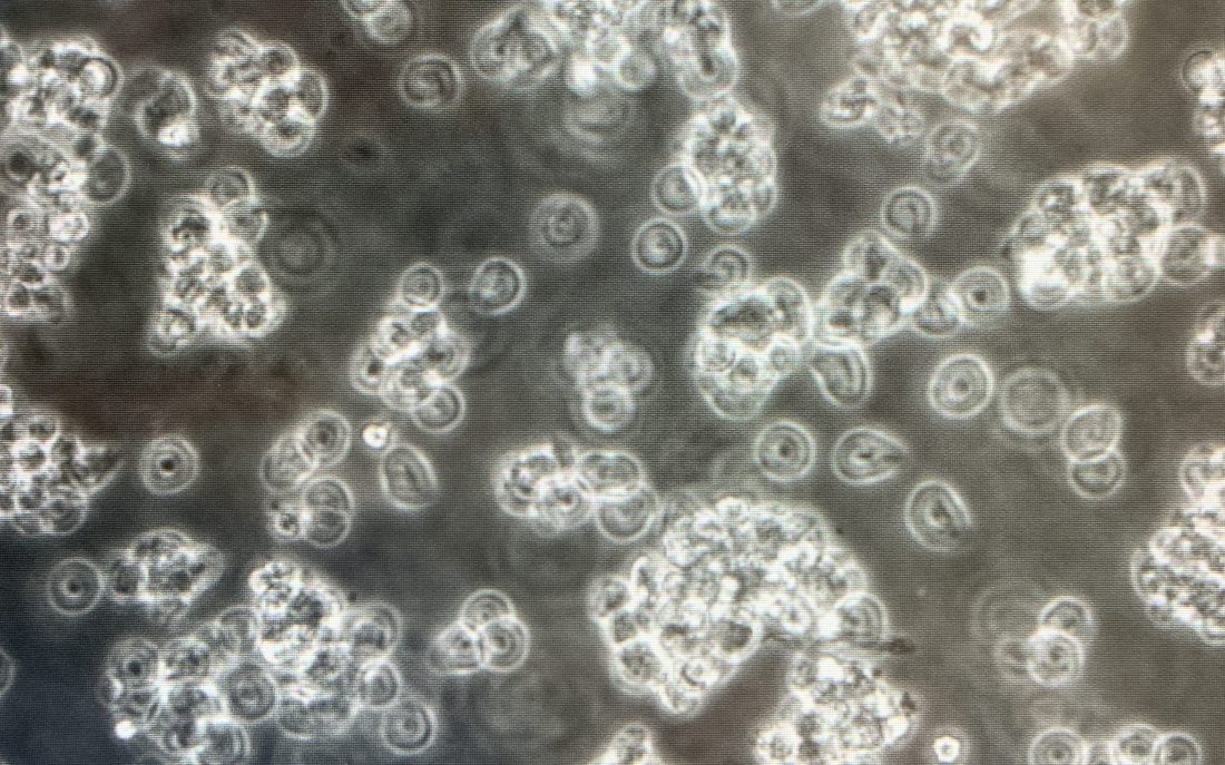
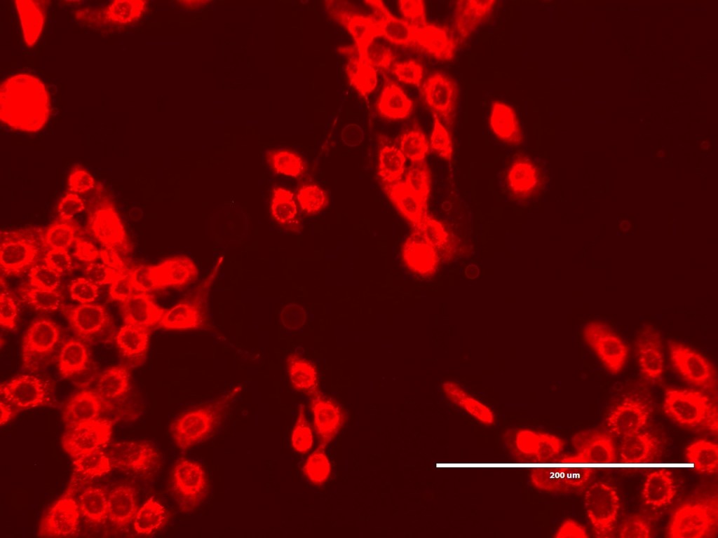
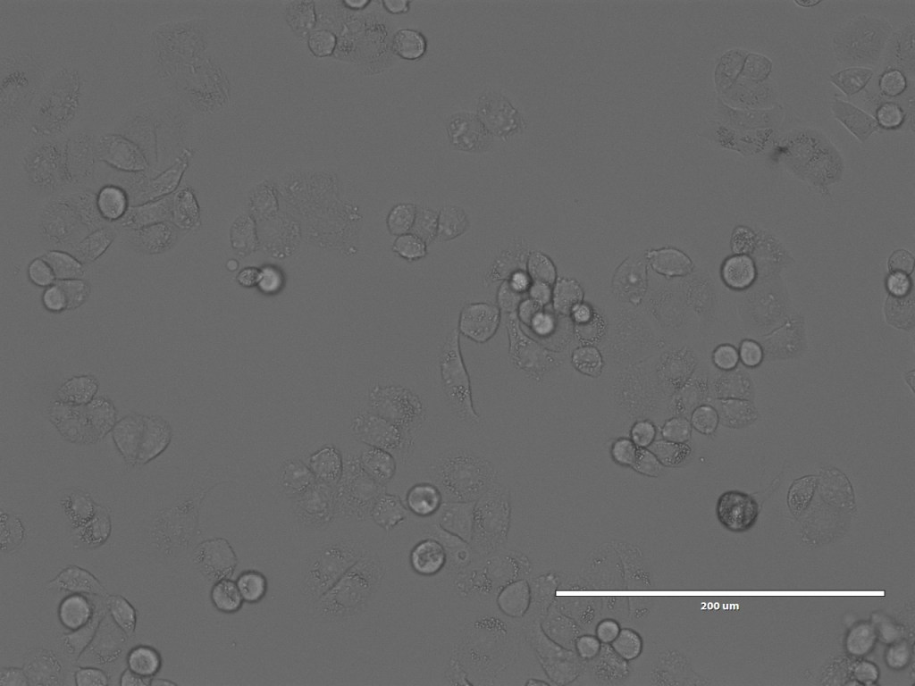
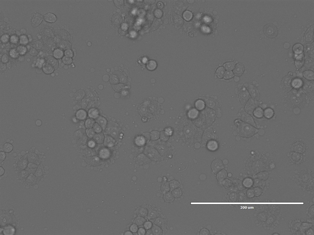
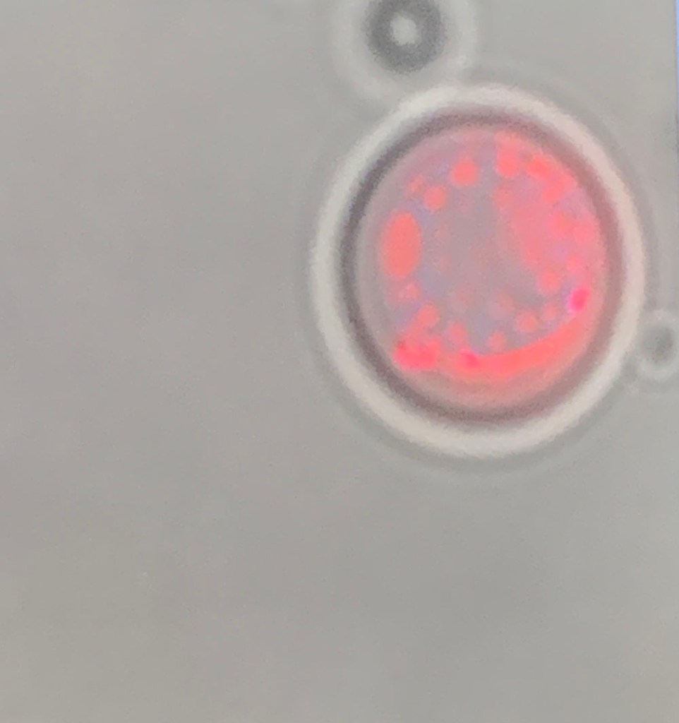
Nonetheless, we spent a happy hour looking at the GPMVs that she had created from the different cell lines. One of the interesting things for me was not only to see the GPMVs themselves, but also to see the very different appearances of the different cell lines under the microscope.
The mcf10 cell line GPMVs fluorescing through the microscope along with the cells themselves – the GPMVs are the rather fainter spheres,Au565 cells and GPMV under normal lightThe 468 cell line and GPMVs under normal light
So that brings us more or less to the end of the trip. I did also spend a bit of time with Chris and Debbie in the Bexley Wing and Clinical Research Facility looking at the potential spaces for the piece, but will post separately about that.
Overall it was a wonderful, stimulating visit. I am still bursting with ideas for tests and approaches to making the artwork, even though everything has been disrupted by the Coronavirus spread and the lockdown, which, frankly, has made it much harder to focus on this, or indeed anything.
If you haven’t seen my post about my first day visiting the labs, it’s probably worth starting there. If you have, welcome to day two!
On my second day I went back to meet Andrew at the School of Chemistry. We chatted while he prepared a lipid mixture to ‘grow’ some GUVs (Giant Unilamellar Vesicles). He prepared two mixtures. One he knew would make it quite tricky to grow the GUVs but would give us the chance to run a new experiment if we got good GUVs, the other was a more reliable but less exciting mix so that we would have something to look at through the confocal microscope later if the first mix failed.
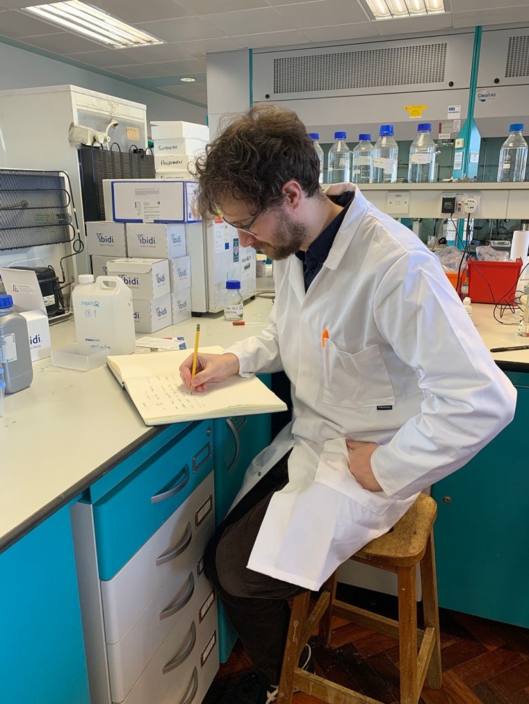
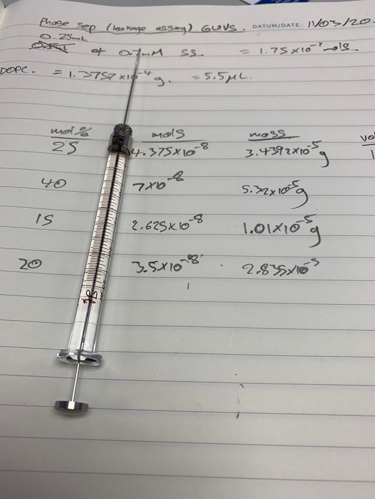
Andrew making calculations
Making GUVs is not done using the same technique as preparing Large vesicles (LUVs), which is what Luke, a Masters student in the lab, was involved in, alongside us in the lab. (The ‘Giant’ and the ‘Large’ are all relative of course – we are talking microns here). He recounted how he had been trying for months to create GUVs of a certain type and over those several months it had only worked well once – on a day when, atypically, he didn’t have the info to replicate the conditions. I compared that to trying to repeat firings to try and find a good programme for my pate de verre sacks – my Imperfect Vessels – which in the end took about 12 firings to get a programme that worked the way I wanted it to. That conversation made me feel a good deal less patient or diligent than I had.
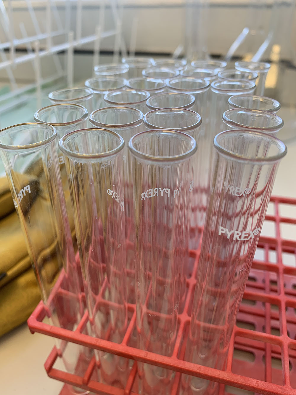
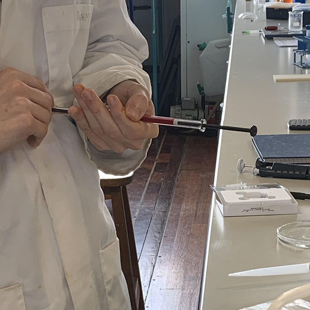
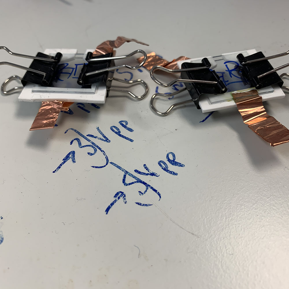
Luke ‘extruding’ his LUVsOne of the teeny tiny syringes used to measure out the right amount of each of the lipids – the central channel that draws up the liquid is barely the width of a wire. I loved them.The lipid mixture is placed on a glass plate with one conductive side and then paired with another plate separated silicon. Once held with bulldog clips (so useful those things) they are connected up and put into the oven, which is stepped slowly to process temperature. Bit like a kiln.
Having wired up the GUVs and popped them into the oven to form, we went to look at the chemical robot. This is a machine for setting up plates with multiple samples at a time according to whatever is programmed for a specific set of experiments. It was strangely mesmerising – whoever had developed the machine had programmed in a set of elegant flourishes – not obviously functional, but very engaging. As the pipettes dipped towards the plates they each dropped down in turn, creating a lovely wave of pipettes heading into the wells with their samples. Similarly, there was a moment’s dramatic pause before the used pipette heads were all knocked off and the sequence started again. A kind of magic.
Some shaky video of the chemical robot doing its dance
In a quiet moment between experiments etc, we started talking about the Brazilian wasp from which the MP1 peptide is drawn. It’s called Polybia Paulista and it’s massive!
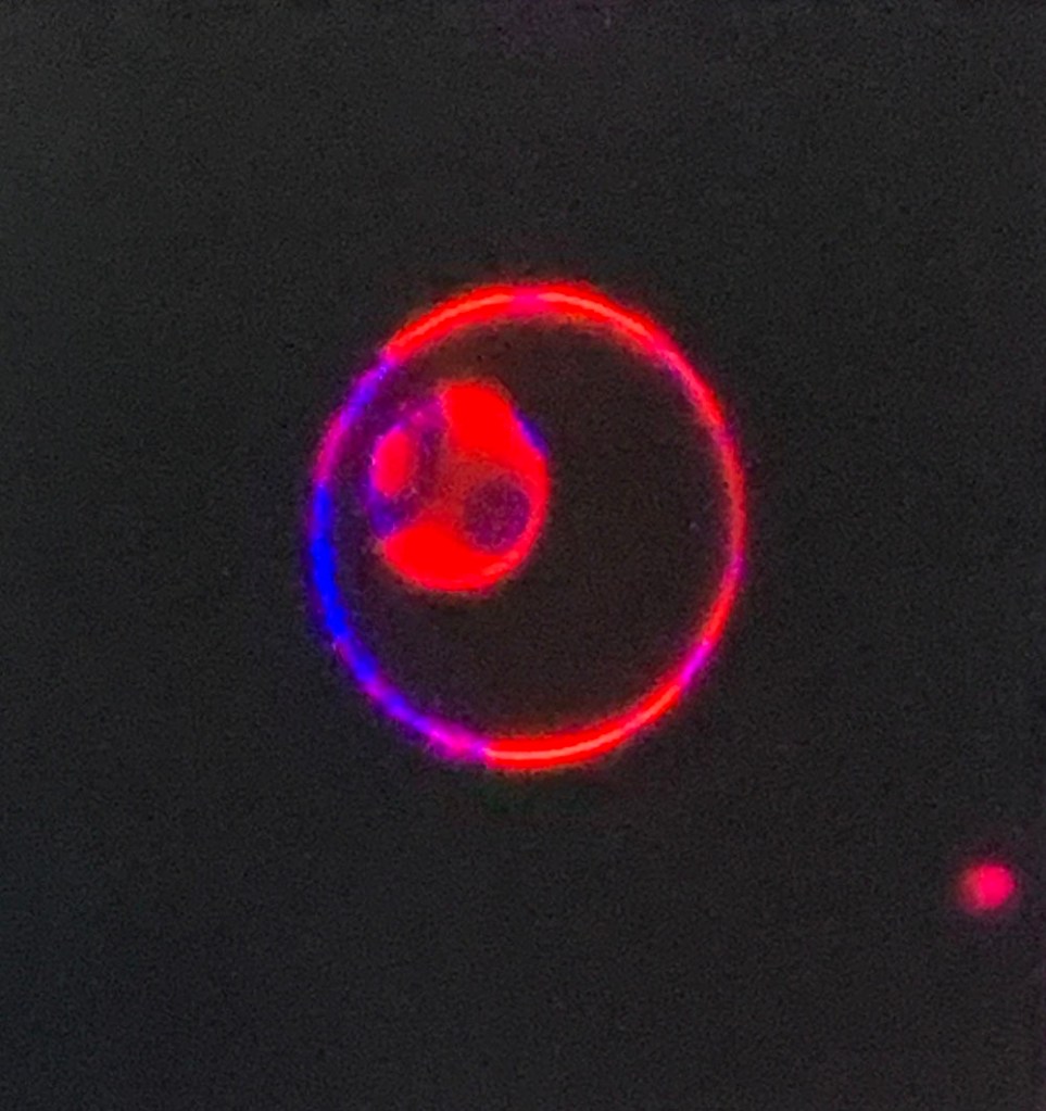
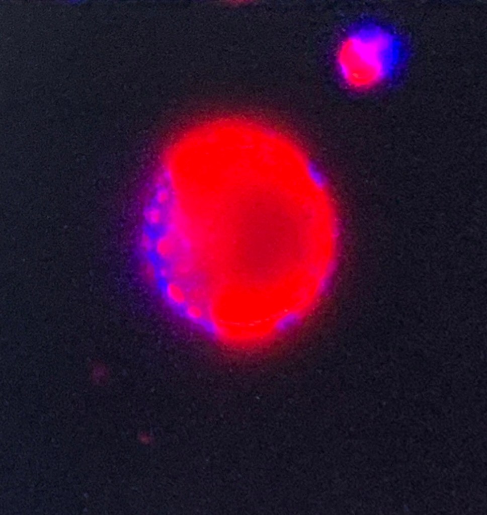
Later in the afternoon we took a look at our GUVs through the confocal microscope. Here are some of the images I snapped off the screen
The images are super-seductive, with the bright coloured labelling (this is showing phase separation – more of that later). But although the temptation is to leap into making bright, spotty, luminescent glass forms, I am not sure that’s where i should ultimately be headed.
In the evening, Paul and I continued the conversations over dinner, and then met up with some of his colleagues from The Superposition, an amazing art/science collective in Leeds. It was a lovely evening and brilliant to chat, but everyone’s anxiety about Coronavirus was already surfacing, and i headed off feeling a bit sombre.
So, last week I travelled up to Leeds to visit the team doing the research into membrane disrupting peptides that is behind my commission. This post is really a brief skip across my time spent in the labs, the basic narrative of the three days spent in the lab environment. There will be other posts about some of the conversations, some of my impressions and my thinking as it develops – in these days of Coronavirus isolation, I am taking my time to process my thoughts – it appears there’s no rush at the moment so I am going to make the most of that!
I arrived at the Uni around midday to be met by Paul, and we had an hour or so to catch up before he had organised a group meeting so i could get to know the rest of the team and start to try and understand the project, the research and the basics of the science behind what they are doing. Before I go any further, I should say that everyone was welcoming, super-patient with all my (often daft) questions and bent over backwards to help me understand and be inspired. Everything that follows in this post (and others) is my interpretation of what I learned, and I must say that any mistakes or strange extrapolations are all mine.
Can you see me?
Over a sandwich, Paul took me through some of the very basics about the science of membranes and the chemistry of lipids. We talked about morphology, hydrophobia and hydrophilia, and heads and tails. We wandered through the basics of peptides, the specific peptide they are working with (which I now know as MP1) and peptide variants. We talked about the effects of soap on membranes – super topical in the developing Coronavirus context. And then we went to the group meeting.
Dyes and Lipids
The group working on the project on a day to day basis is the coming together of two perspectives. Paul and Andrew, based in the School of Chemistry, come from what I think of as the ‘molecular point of view’, focusing on the behaviour of the lipids, peptides structurally and chemically. Tom and Arindam focus on the ‘biological perspective’, working with cell lines and 3D cell clusters (spheroids and similar), testing the peptide on live cells rather than vesicles created out of specific cocktails of lipids. That’s actually quite a simplistic way to look at it, but at present it provides me with a mental framework that sparks ideas. But more of that later.
That evening, Tom and I went out to eat pasta and chat about science, art, northern towns, biology and the funding of scientific journals – a bleedin’ shocker as far as I’m concerned, which I am sure I shall come back to in due course. If anyone is interested in some of the background in the meantime though, I would recommend reading this Guardian Long Read about the history and development of science publishing.